Spinal Fusion Devices: A Complete Guide for Surgical Teams
Spinal fusion devices are the hardware and biological materials that hold the spine rigid while bone grows across the treated motion segment, permanently eliminating movement at that level. The construct has two jobs: provide immediate mechanical stability (the hardware) and create the biological conditions for bone fusion (the biologics). If the hardware fails before fusion consolidates, the case fails. If the biology fails and fusion never occurs, the hardware will eventually fail from fatigue. Both halves of the equation must work.
This guide covers every major category of spinal fusion device in current surgical use: pedicle screw and rod systems, interbody fusion cages across all surgical corridors, cervical plating, supplemental fixation, and the biologics that drive the fusion process. It is built for spine surgeons, surgical technologists, device representatives, and procurement teams who need to understand what each device does, when it is indicated, and how these components work together as a system.
Principles of Spinal Fusion
Spinal fusion (arthrodesis) is the biological process of growing bone across a motion segment to eliminate movement. The goal is not just hardware placement — it is solid bony union between adjacent vertebral bodies (interbody fusion), between transverse processes (posterolateral fusion), or both (360-degree or circumferential fusion).
For fusion to succeed, three conditions must be present — often called the “diamond concept” of bone healing:
- Mechanical stability — the instrumentation must immobilize the segment sufficiently to allow bone to bridge without excessive micromotion disrupting the healing process. This is the job of the pedicle screws, rods, interbody cages, and plates.
- Biological environment — the fusion bed must contain osteogenic cells (cells that form new bone), osteoinductive signals (growth factors that recruit and stimulate bone-forming cells), and an osteoconductive scaffold (a structural matrix for new bone to grow on and through).
- Adequate vascularity — blood supply to the fusion bed delivers oxygen, nutrients, and progenitor cells. Surgical technique that preserves soft tissue attachments and blood supply promotes fusion. Excessive muscle stripping and devascularization of the bone surfaces impair it.
The devices covered in this guide address the first two conditions. The third is a function of surgical technique.
Pedicle Screw and Rod Systems
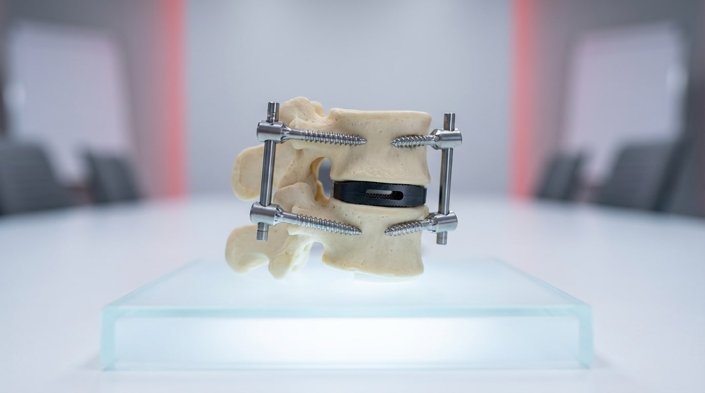
Pedicle screw fixation is the backbone of posterior spinal fusion. A screw is placed through the pedicle of each vertebra into the vertebral body, and bilateral screws at adjacent levels are connected by rods. The resulting construct immobilizes the instrumented segments in all planes — flexion/extension, lateral bending, and rotation.
Screw Types
- Polyaxial screws — the screw head pivots in multiple directions (typically 25-30 degrees of freedom) before the rod is locked in the saddle. This angulation freedom makes rod-screw coupling easier across multiple levels, particularly when pedicle anatomy varies or the spine has deformity. Polyaxial screws are the standard for most fusion cases.
- Monoaxial screws — fixed-head design with no angulation between the screw shaft and the rod capture mechanism. Monoaxial screws provide maximum rigidity and are used primarily in fracture fixation and deformity correction where controlled correction forces must be transmitted through the screw without any angular play.
- Reduction screws — polyaxial screws with extended tabs that allow sequential rod reduction. In spondylolisthesis or kyphotic deformity, the rod sits above the screw head and must be reduced (brought down) into the saddle. Reduction screws provide the mechanical advantage to accomplish this without forcing the anatomy.
- Cannulated screws — hollow-core screws placed over a guidewire under fluoroscopic guidance. Standard for percutaneous (minimally invasive) pedicle screw placement. The guidewire is placed first, trajectory is confirmed on imaging, and the screw is advanced over the wire.
- Fenestrated screws — screws with side holes along the shaft for PMMA cement augmentation in osteoporotic bone. After the screw is placed, cement is injected through the cannulation and extrudes through the fenestrations into the surrounding cancellous bone, creating a cement mantle that increases pullout strength by 100-200%.
Rod Systems
Connecting rods are available in titanium alloy (Ti-6Al-4V) and cobalt-chrome (CoCr). Titanium rods are the standard for most fusion cases — they offer adequate stiffness, fatigue resistance, and MRI compatibility. CoCr rods provide approximately twice the stiffness of titanium and are preferred for long-segment deformity constructs where greater corrective force and resistance to rod fracture are needed.
Standard rod diameters are 5.5mm and 6.0mm for the thoracolumbar spine, and 3.5mm for the cervical spine. Pre-contoured rods (with lordotic or kyphotic curves built in) reduce intraoperative bending and provide a more anatomic fit. Straight rods require intraoperative contouring with rod benders — a process that introduces stress risers at the bend points and can reduce fatigue life if not performed carefully.
Set Screws and Locking Mechanisms
The set screw is what locks the rod into the screw head. Modern systems use top-loading set screws with a defined break-off torque (typically 8-12 Nm) that ensures consistent locking force without over-tightening. Some systems use side-loading or bottom-loading rod capture mechanisms, each with different advantages for rod insertion in tight surgical corridors (MIS cases, revision surgery with scar tissue).
Interbody Fusion Cages
Interbody cages are structural devices placed in the disc space after discectomy to restore disc height, maintain foraminal height (decompressing the exiting nerve root), and provide a scaffold for bone graft material that will achieve interbody fusion. The cage bears axial load while fusion consolidates.
Cages are classified by the surgical corridor used to access the disc space. For more detail on the instrumentation used in each approach, see our spine surgery instrumentation guide.
ALIF (Anterior Lumbar Interbody Fusion) Cages
Placed through an anterior (abdominal) approach. ALIF cages are the largest interbody devices, offering the greatest footprint on the vertebral endplate and the largest volume for bone graft. They are typically standalone devices with integral fixation (screws through the cage into the adjacent vertebral bodies) or used with supplemental posterior fixation. ALIF cages provide excellent lordosis restoration and indirect decompression through disc height restoration.
PLIF (Posterior Lumbar Interbody Fusion) Cages
Placed through a posterior approach after bilateral laminectomy and facetectomy. Two smaller cages are placed side by side in the disc space (one on each side of the thecal sac). PLIF provides both direct posterior decompression and interbody fusion in a single approach but requires retraction of the neural elements, which carries a risk of dural tear and nerve root injury.
TLIF (Transforaminal Lumbar Interbody Fusion) Cages
Placed through a posterolateral approach via unilateral facetectomy. The cage enters the disc space from the side, reducing the need to retract neural structures. TLIF has become the most commonly performed interbody fusion technique because it provides interbody fusion with less neural retraction than PLIF and can be performed through minimally invasive approaches. TLIF cages are available in static (fixed height and lordosis) and expandable (adjustable height after insertion) designs.
LLIF / XLIF (Lateral Lumbar Interbody Fusion) Cages
Placed through a lateral (transpsoas or anterior-to-psoas) approach. Lateral cages have a large footprint and span the full width of the disc space, resting on the strong peripheral ring apophysis of the vertebral endplate rather than the weaker central endplate. This produces excellent indirect decompression, lordosis correction, and coronal plane deformity correction. Lateral approaches avoid the morbidity of anterior abdominal surgery and the neural retraction of posterior approaches. The trade-off is the risk of lumbar plexus injury (psoas-related neuropraxia), which is approach-specific.
Cage Materials
- PEEK — radiolucent, modulus close to bone, allows clear radiographic fusion assessment. No bone ingrowth capability — fusion occurs through the graft material inside the cage, not through the cage walls.
- Titanium (solid or 3D-printed) — radiopaque, higher modulus than PEEK, superior osseointegration. 3D-printed titanium cages with open-lattice porous surfaces allow bone to grow through the cage structure itself, creating biological fixation at the cage-endplate interface in addition to interbody fusion through the graft window.
- Porous tantalum — high porosity (75-80%), modulus close to cancellous bone, excellent ingrowth properties. Used in some interbody cage designs.
- Biocomposite — resorbable polymers combined with calcium phosphate ceramics. Gradually resorbed and replaced by bone. Limited clinical adoption in interbody applications due to concerns about load-bearing capacity during the resorption phase.
Static vs. Expandable Cages
Static cages are inserted at their final height and lordosis. The surgeon selects the appropriate trial size and inserts the corresponding implant. Expandable cages are inserted at a reduced height and then expanded in situ using a mechanical actuator. The advantage is the ability to insert through a smaller corridor (important for MIS TLIF and lateral approaches) and then dial in the exact height and lordosis after the cage is seated. The potential disadvantage is mechanical complexity — more moving parts mean more potential failure modes over time.
Cervical Plating Systems
Anterior cervical plates are used in ACDF (anterior cervical discectomy and fusion) and corpectomy procedures to stabilize the cervical spine after disc or vertebral body removal. The plate spans the fused segment, with screws placed into the vertebral bodies above and below the graft or cage.
Plate Design Features
- Fixed-angle screws — screws lock into the plate at a fixed trajectory, providing rigid fixation. Used in fracture and corpectomy constructs where immediate stability is critical.
- Variable-angle screws — screws can be angled within a cone of freedom (typically 12-15 degrees), allowing the surgeon to adjust trajectory to avoid adjacent endplates or to optimize purchase in the vertebral body. Most modern cervical plates offer variable-angle screw options.
- Dynamic plates — plates that allow controlled settling of the graft or cage as it is loaded during healing. A small amount of axial shortening is permitted through a sliding mechanism in the plate, which maintains compressive load on the graft and theoretically promotes fusion. Excessive settling can result in loss of disc height and foraminal stenosis.
- Semi-constrained plates — plates that allow some screw toggling or rotation after locking, providing a middle ground between rigid and dynamic constructs.
Low-Profile and Zero-Profile Designs
Standard anterior cervical plates sit on the anterior surface of the vertebral bodies and are covered by the prevertebral soft tissues. Plate prominence can cause dysphagia — difficulty swallowing caused by the plate pushing against the esophagus. Low-profile plates minimize anterior projection. Zero-profile devices integrate the fixation screws directly into the interbody cage, eliminating the anterior plate entirely. These cage-with-integrated-screws designs have been shown to reduce dysphagia rates compared to standard plate-and-cage constructs.
Supplemental Fixation: Cross-Connectors, Hooks, and Wires
Cross-Connectors
Cross-connectors (transverse connectors) link the bilateral rods, converting them from two parallel beams into a frame. This significantly increases the construct’s resistance to axial rotation. Cross-connectors are standard in multi-level posterior fusions (three or more levels) and deformity constructs. In single-level or two-level fusions, the rotational stiffness provided by bilateral pedicle screws and an interbody cage is typically sufficient without a cross-connector.
Hooks and Wires
Laminar hooks and sublaminar wires were the primary fixation methods before pedicle screws became standard. They are now used as supplemental fixation in specific situations: proximal hooks at the top of a long deformity construct to reduce the risk of proximal junctional failure, sublaminar bands (polyester or titanium cable) for coronal plane correction in scoliosis, and as salvage fixation when pedicle anatomy does not permit screw placement.
Sacropelvic Fixation
Long constructs extending to the sacrum require sacropelvic fixation to anchor the caudal end of the construct. S2-alar-iliac (S2AI) screws have largely replaced traditional iliac screws. S2AI screws are placed through the S2 pedicle, across the sacroiliac joint, and into the ilium — all in-line with the pedicle screw system, eliminating the need for offset connectors. They provide excellent fixation strength and lower implant prominence compared to iliac screws.
Biologics for Spinal Fusion
Hardware provides stability. Biologics provide fusion. Every spinal fusion construct requires a biological strategy to achieve bony union. The choice of biologic material is as consequential as the choice of hardware.
Local Autograft
Bone harvested during the decompression (lamina, spinous process, facet) is the gold standard graft material. It contains viable osteogenic cells, osteoinductive growth factors, and an osteoconductive scaffold — all three elements of the fusion biology. The limitation is volume: in a minimally invasive case or a single-level ACDF, the available local bone may be insufficient.
Iliac Crest Bone Graft (ICBG)
Autograft harvested from the iliac crest provides a larger volume than local bone. It was the historical gold standard but has declined in use due to donor site morbidity — pain, hematoma, sensory nerve injury, and cosmetic deformity at the harvest site. ICBG is still used in complex revision cases, pseudarthrosis repair, and situations where maximum biological potency is needed.
Allograft
Cadaveric bone (cortical, cancellous, or corticocancellous) provides an osteoconductive scaffold and weak osteoinductive signaling but no living cells. Structural allograft (femoral ring, machined cortical spacers) is used as an interbody spacer in ACDF. Cancellous allograft chips are used as graft extenders in posterolateral fusion beds, mixed with local autograft to increase graft volume.
Demineralized Bone Matrix (DBM)
Processed allograft with the mineral content removed, exposing the collagen matrix and endogenous growth factors. Available as putty, gel, strips, and fiber. DBM is a graft extender — it adds osteoinductive and osteoconductive properties to a graft composite but is not sufficient as a standalone graft material for interbody fusion.
Cellular Allografts
Products containing viable mesenchymal stem cells or osteoprogenitor cells from amniotic, umbilical, or placental tissue. These add the osteogenic component that standard allograft and DBM lack. Clinical adoption is growing, though the evidence base is still maturing. Combining a cellular allograft with local autograft and an osteoconductive scaffold creates a composite strategy that addresses all three biological pillars.
Bone Morphogenetic Protein (BMP-2)
Recombinant human BMP-2 (INFUSE, Medtronic) is the most potent osteoinductive agent available. FDA-approved for ALIF with a specific cage. Off-label posterior use has decreased due to complications including ectopic bone formation, radiculitis, and osteolysis. Cervical use is associated with airway swelling and is generally contraindicated. BMP-2 remains useful in specific clinical scenarios with appropriate informed consent and evidence-based dosing.
Synthetics
Beta-tricalcium phosphate (TCP), hydroxyapatite (HA), and biphasic ceramics serve as osteoconductive scaffolds. They are graft extenders — combined with biologically active materials to increase total graft volume. They do not have osteoinductive or osteogenic properties independently.
Device Selection: Matching Hardware to the Clinical Problem
The device selection decision is not about choosing the newest or most expensive implant. It is about matching the mechanical requirements of the clinical problem to the capabilities of the hardware and biologics.
- Single-level degenerative disc disease with stenosis — TLIF cage + bilateral pedicle screws and rods, with local autograft + DBM or cellular allograft. This is the workhorse construct.
- Cervical disc herniation with radiculopathy — ACDF with interbody cage (PEEK or titanium) + anterior cervical plate or zero-profile integrated device, with allograft or DBM in the cage.
- Two-level spondylolisthesis — bilateral pedicle screws with reduction screws at the listhetic level, TLIF or PLIF cages at both levels, CoCr rods if significant reduction force is needed.
- Adult degenerative scoliosis — long-segment posterior instrumentation (pedicle screws at all levels), lateral interbody cages at multiple levels for coronal and sagittal correction, CoCr rods, S2AI screws, cross-connectors, and an aggressive biologic strategy (local autograft + BMP or cellular allograft at every fusion level).
- Osteoporotic compression fracture with kyphosis — short-segment pedicle screw fixation with fenestrated cement-augmented screws, possible vertebral body augmentation (kyphoplasty), and titanium rods.
Supply Chain Considerations
Spinal fusion cases are instrument-intensive. A single posterior lumbar fusion requires a pedicle screw tray, rod tray, cage tray with trials, and potentially a biologics delivery system. A multi-level deformity case may require four or five instrument trays plus specialized reduction, deformity correction, and navigation instruments. If any tray is missing or incomplete, the case is compromised or cancelled.
This makes supplier reliability the most operationally critical factor in spine device procurement. A supplier with the right devices but the wrong delivery model — one that requires 72-hour lead time, ships from a distant distribution center, or cannot guarantee complete tray sets — creates recurring operational risk for every case on your schedule.
SLR Medical Consulting supplies spinal fusion devices, orthopedic hardware, and biologics to surgical facilities nationwide from fully stocked warehouses with zero-lead-time processing. Complete tray sets, delivered on your schedule. Browse our spine hardware catalog or place a surgical order.
Frequently Asked Questions About Spinal Fusion Devices
What is the difference between a TLIF and an ALIF cage?
A TLIF cage is inserted from a posterolateral approach (through a facetectomy) and is typically kidney-bean or banana-shaped to fit through the narrow corridor between the thecal sac and the pedicle. An ALIF cage is inserted from an anterior (abdominal) approach and has a much larger footprint — it spans the full anterior disc space. ALIF cages provide greater lordosis correction and more bone graft volume but require an anterior surgical approach (with vascular risk). TLIF cages can be placed through the same posterior incision used for pedicle screw placement, avoiding a separate anterior procedure. The choice depends on the pathology, the amount of lordosis correction needed, surgeon training, and whether a circumferential fusion is planned.
When should cement-augmented pedicle screws be used?
Cement-augmented (fenestrated) pedicle screws should be considered when bone density is significantly reduced and standard screw fixation is at risk of pullout failure. Practical indicators include patients over 65 with known osteoporosis, DEXA T-scores below -2.5, vertebral compression fractures indicating poor bone quality, and revision cases where prior screw tracts have enlarged. Cement augmentation increases pullout strength by 100-200% but carries risks including cement leakage into the spinal canal or foramen, and embolization. Careful technique with controlled injection under fluoroscopic guidance minimizes these risks.
What biologics should be used for a standard single-level posterior lumbar fusion?
For a standard single-level TLIF, the most common biologic strategy combines local autograft (bone harvested from the laminectomy and facetectomy) with a graft extender such as demineralized bone matrix (DBM) or a cellular allograft. Local autograft is packed inside the interbody cage to promote interbody fusion, and the remaining local bone plus DBM is placed in the posterolateral gutter. This composite approach addresses all three biological requirements: osteogenic cells (from the autograft), osteoinductive signals (from the DBM and autograft matrix), and an osteoconductive scaffold (the cage and the allograft/DBM carrier). BMP-2 is an option but carries cost and complication considerations that must be weighed against the clinical scenario.
How do 3D-printed titanium cages differ from PEEK cages clinically?
3D-printed titanium cages have an open-lattice porous surface that promotes bone ingrowth directly into the cage structure, creating biological fixation at the cage-endplate interface. PEEK cages are bioinert — bone does not grow into PEEK. Fusion with PEEK cages occurs through the bone graft inside the cage, not through the cage material itself. Titanium cages produce imaging artifact on CT and MRI, making radiographic fusion assessment harder. PEEK is radiolucent, allowing clear visualization of fusion progress. Early clinical data suggest comparable fusion rates between modern 3D-printed titanium and PEEK cages, though some series report faster radiographic fusion with porous titanium designs. The choice often depends on surgeon preference and whether postoperative imaging clarity is a priority for the specific case.
About SLR Medical Consulting: SLR Medical Consulting has been supplying surgical facilities nationwide for over a decade with orthopedic hardware, spine instrumentation, biologics, and sports medicine devices. Our zero-lead-time delivery model means your surgical schedule runs on your timeline, not your supply chain’s. Explore our hardware catalog or place a surgical order today.