The Evolution of Spine Surgery Technology
Spine surgery has undergone more technological transformation in the last sixty years than almost any other surgical specialty. A patient undergoing a lumbar fusion in 1965 faced a fundamentally different operation — different instrumentation, different fixation, different recovery timeline, different risk profile — than a patient undergoing the same diagnosis-driven procedure today. The implants are different. The approaches are different. The imaging is different. The outcomes data is incomparable.
But the evolution didn’t happen in a straight line. It moved in waves, each one driven by a specific engineering breakthrough or clinical insight that changed what surgeons could do inside the spinal column. Some of those breakthroughs held up. Some created problems that took a decade to recognize. Understanding this history matters if you work in the spine device space — because the technology decisions being made in operating rooms today are direct descendants of innovations that started in the 1950s and 1960s.
This article traces the major inflection points in spine surgery technology, from the earliest instrumentation systems through the robotic-assisted minimally invasive procedures being performed right now.
The Early Era: Harrington Rods and the Birth of Spinal Instrumentation
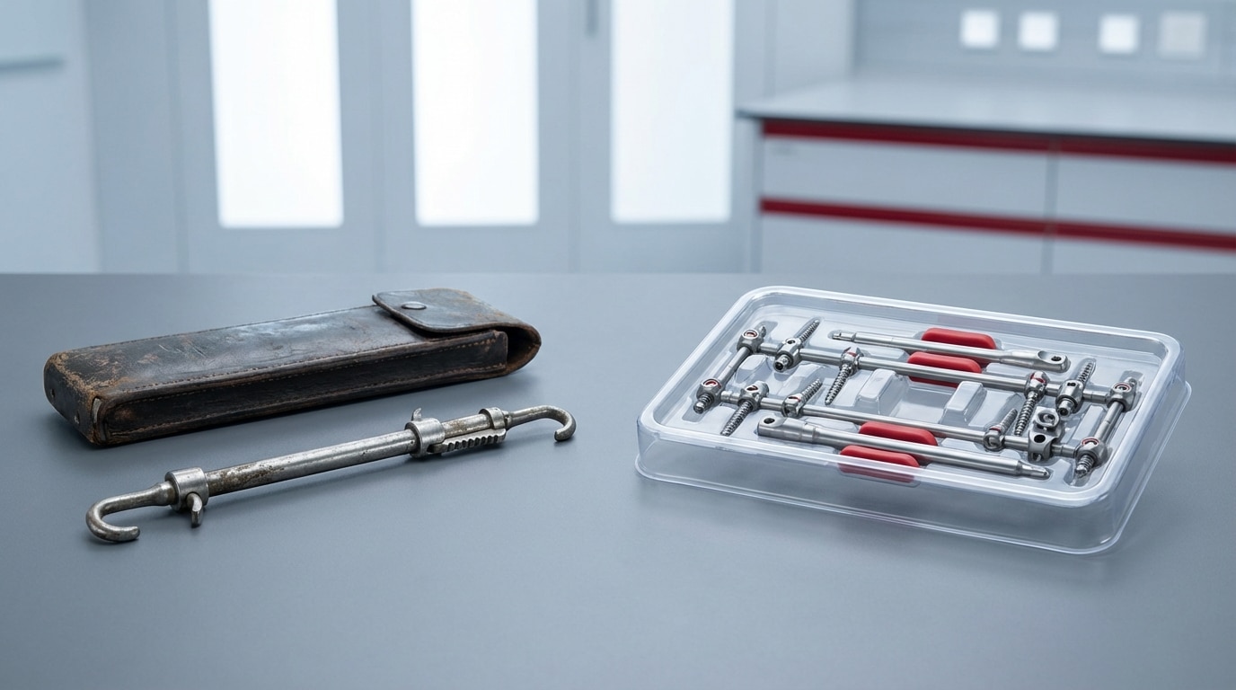
Before Paul Harrington developed his distraction rod system in the 1950s and 1960s, spinal surgery for scoliosis and fractures was largely limited to posterior fusion without internal fixation. Surgeons would decorticate the posterior elements, lay down bone graft, and hope the fusion consolidated while the patient spent months in a body cast or brace. The nonunion rates were high. The correction was unpredictable. The patient experience was brutal.
Harrington’s system used stainless steel rods with ratcheted hooks that could be distracted along the posterior spine to correct curvature. For scoliosis patients, this was transformative. A severe curve that would have required prolonged casting could now be partially corrected intraoperatively and held in position while fusion occurred. The Harrington rod became the standard of care for scoliosis surgery for nearly two decades.
But the system had real limitations. Harrington rods provided distraction — they could lengthen the concavity of a curve — but they couldn’t address rotation, which is a fundamental component of scoliotic deformity. They also required long fusions, often extending well below the curve to maintain balance, which sacrificed lumbar motion segments that didn’t need to be fused. The phenomenon of “flatback syndrome,” where loss of lumbar lordosis from long Harrington rod constructs caused patients to pitch forward, became a recognized complication that surgeons dealt with for years after the initial procedure.
Despite these problems, the Harrington rod era established a critical principle: internal fixation could improve spinal surgery outcomes. That principle drove everything that came after.
Pedicle Screws: The Innovation That Changed Everything
The introduction of pedicle screw fixation in the 1980s was the single most important technical development in the history of spine surgery. Nothing else comes close in terms of impact on what procedures became possible and how reliably they could be performed.
Roy-Camille in France and Magerl in Switzerland were among the pioneers who demonstrated that screws could be safely placed through the pedicles of the vertebrae into the vertebral body. This provided three-column fixation — the screw engaged the posterior elements through the pedicle and anchored in the anterior vertebral body — which was biomechanically superior to any hook or wire-based system.
The clinical impact was immediate and measurable:
- Segmental fixation. Unlike Harrington rods that spanned long segments with hooks at the ends, pedicle screws could be placed at every level being fused, providing segmental control of each vertebra. This allowed shorter fusion constructs and better correction of deformity.
- Three-dimensional correction. Pedicle screws could address translation, rotation, and angulation — all three planes of spinal deformity. Harrington rods primarily addressed the coronal plane.
- Improved fusion rates. The rigid fixation provided by pedicle screws created a more stable mechanical environment for bone healing, which translated directly into higher fusion rates and lower pseudarthrosis rates.
- Enabled new procedures. Posterior lumbar interbody fusion (PLIF) and later transforaminal lumbar interbody fusion (TLIF) became practical procedures because pedicle screws provided the posterior stabilization needed to support interbody grafts.
The FDA controversy around pedicle screws is worth noting because it shaped the regulatory environment for spine devices going forward. Pedicle screws were used widely throughout the 1980s and early 1990s as an off-label application of bone screw technology. The FDA held an advisory panel in 1994 that raised safety concerns, leading to a period of regulatory uncertainty. Eventually, Class II clearance pathways were established, but the episode highlighted the tension between clinical adoption and regulatory oversight that still exists in the spine device market.
By the mid-1990s, pedicle screw systems from companies like Synthes, DePuy, Sofamor Danek (later acquired by Medtronic), and Stryker had become the backbone of posterior spinal instrumentation. Today, it is nearly impossible to imagine spine surgery without them.
Cages and Interbody Fusion: Rethinking the Anterior Column
The development of interbody fusion devices — cages — addressed a problem that pedicle screws alone couldn’t solve: restoring disc height, achieving anterior column support, and creating an optimal environment for fusion across the disc space.
The Bagby and Kuslich (BAK) cage, developed in the early 1990s, was among the first FDA-cleared interbody devices. It was a threaded cylindrical titanium cage that was packed with bone graft and inserted into the disc space after discectomy. The concept was straightforward: the cage maintained disc height and foraminal dimensions while the bone graft inside and around it consolidated into a solid fusion.
From that starting point, interbody technology branched rapidly:
- PEEK cages replaced titanium in many applications because PEEK (polyetheretherketone) has a modulus of elasticity closer to bone, reducing stress shielding, and it is radiolucent, allowing surgeons to visualize bone graft consolidation on X-rays and CT without metal artifact.
- Expandable cages allowed surgeons to insert a smaller-profile device through a minimally invasive corridor and then expand it in situ to restore disc height and lordosis. This was a critical enabling technology for lateral approaches.
- 3D-printed titanium cages with porous surface architectures designed to mimic trabecular bone structure emerged in the 2010s. The theory is that bone cells can grow into the porous lattice, creating a direct bone-implant interface rather than relying solely on bone graft within and around the cage.
- Lordotic and hyperlordotic designs gave surgeons the ability to restore sagittal alignment through the cage geometry itself, which became increasingly important as the spine surgery field focused more on sagittal balance as a predictor of clinical outcomes.
The approach corridors for interbody fusion also multiplied. ALIF (anterior lumbar interbody fusion), PLIF, TLIF, LLIF/XLIF (lateral lumbar interbody fusion), and OLIF (oblique lateral interbody fusion) each offer different access points to the disc space with different risk profiles and different cage geometries optimized for each approach.
For device sales professionals working in spine, understanding cage options and approach corridors is table-stakes knowledge. Surgeons have strong preferences, and those preferences are often tied to their training, their complication experience, and the specific pathology they’re treating.
The MIS Revolution: Smaller Incisions, Bigger Questions
Minimally invasive spine surgery (MIS) began gaining serious traction in the early 2000s, driven by the same forces that pushed MIS adoption across all surgical specialties: less tissue disruption, less blood loss, shorter hospital stays, and faster return to function.
The key enabling technologies for MIS spine surgery were tubular retractors and percutaneous pedicle screw systems. Tubular retractors allowed surgeons to perform decompressions and interbody fusions through small incisions by dilating the paraspinal muscles rather than stripping them from the bone. Percutaneous screw systems allowed pedicle screws to be placed through small stab incisions under fluoroscopic guidance, avoiding the extensive posterior exposure required for traditional open screw placement.
The MIS approach delivered real benefits in soft-tissue preservation. Studies consistently showed less blood loss, shorter hospital stays, and lower infection rates compared to open surgery for similar procedures. For simple decompressions and single-level fusions, the evidence was compelling.
But MIS also introduced trade-offs that the early enthusiasm sometimes obscured:
- Increased radiation exposure. Percutaneous screw placement under fluoroscopy meant more radiation for the patient and, critically, for the surgeon and OR staff. Over a career of MIS cases, the cumulative radiation exposure to the surgeon’s hands and eyes became a legitimate concern.
- Learning curve. Working through a tube with limited visualization is technically demanding. The complication rates during a surgeon’s learning curve for MIS techniques were higher than their open surgery complication rates, which raised questions about the risk-benefit calculation during the adoption phase.
- Limited correction capability. For complex deformity cases requiring significant realignment, the access provided by MIS approaches was often insufficient. Surgeons had to carefully select which cases were appropriate for MIS and which required open techniques.
The current state of MIS spine surgery reflects a maturation of the approach. Experienced MIS surgeons can perform complex multi-level fusions through minimally invasive corridors with outcomes that match or exceed open surgery. But case selection remains critical, and the technology has evolved specifically to address the early limitations — particularly the radiation exposure problem, which is where navigation and robotics entered the picture.
Navigation and Intraoperative Imaging
Intraoperative navigation in spine surgery borrowed concepts from neurosurgery, where frameless stereotaxy had been used for brain tumor localization since the 1990s. The basic principle is the same: register the patient’s anatomy to preoperative or intraoperative imaging, then track instruments in real time relative to that registered anatomy.
The major navigation systems in spine include Medtronic StealthStation, Brainlab, Stryker NAV3i, and Globus ExcelsiusGPS (which combines navigation with robotic guidance). These systems use optical or electromagnetic tracking to show the surgeon where their instruments are relative to the patient’s spine in three dimensions.
The clinical value of navigation is most clearly demonstrated in pedicle screw placement accuracy. Multiple meta-analyses have shown that navigated screw placement has higher accuracy rates than freehand or fluoroscopy-guided techniques, with clinically meaningful reductions in screw misplacement — particularly in the thoracic spine where pedicle anatomy is more complex and the margin for error is smaller.
Intraoperative imaging evolved in parallel. The progression from C-arm fluoroscopy to intraoperative CT (O-arm, Airo, Loop-X) gave surgeons the ability to obtain cross-sectional imaging during the procedure, confirm implant placement before closing, and register navigation systems to the patient’s actual intraoperative anatomy rather than relying on preoperative imaging that might not perfectly match the patient’s positioning on the OR table.
For anyone selling spine instrumentation, the navigation ecosystem is an important part of the conversation. Surgeons who have invested in navigation workflows expect their implant systems to be compatible. Some manufacturers have integrated their implant platforms directly with specific navigation systems, creating a bundled offering that can be difficult for competitors to displace.
Motion Preservation: Disc Replacement and Dynamic Stabilization
The conceptual appeal of motion preservation in the spine is obvious. If the problem is a degenerated disc causing pain, why fuse the segment and transfer stress to adjacent levels when you could replace the disc and maintain motion? Adjacent segment degeneration — the breakdown of levels above or below a fusion — is a real clinical problem, and motion preservation devices were developed specifically to address it.
Cervical disc replacement has been the success story. Devices like the Mobi-C (Zimmer Biomet), Prestige LP (Medtronic), and ProDisc-C (DePuy Synthes) have accumulated long-term data showing non-inferiority or superiority to anterior cervical discectomy and fusion (ACDF) at single and contiguous two-level disease. The FDA has approved several cervical artificial discs, and adoption among fellowship-trained spine surgeons has grown steadily.
Lumbar disc replacement has had a more complicated trajectory. The Charite and ProDisc-L were among the first devices cleared for the lumbar spine, but adoption remained limited compared to cervical. Patient selection criteria are narrower. The surgical approach (anterior) carries vascular risks. And many surgeons remained unconvinced that the long-term motion preservation benefit justified the technical complexity and the risk profile of the anterior lumbar exposure, particularly when TLIF with modern cage designs and biologics was producing reliable results.
Dynamic stabilization — devices designed to control but not eliminate motion — has seen mixed results. The Dynesys system and similar posterior dynamic stabilization devices generated initial enthusiasm but failed to demonstrate clear superiority over fusion in long-term studies. Most spine surgeons have moved away from these devices.
The motion preservation conversation is not over. Next-generation disc replacement designs and improved patient selection criteria may expand the indications. But for now, fusion remains the workhorse procedure in the lumbar spine, and motion preservation is primarily a cervical spine technology with a focused set of indications.
Robotic-Assisted Spine Surgery
Robotic assistance in spine surgery represents the convergence of several technology threads — navigation, intraoperative imaging, and automated instrument guidance — into integrated platforms that aim to improve accuracy, reduce radiation exposure, and enable less invasive approaches.
The current major platforms include:
Globus Medical ExcelsiusGPS. A floor-mounted robotic arm that provides guided trajectories for pedicle screw placement. The surgeon plans screw positions on preoperative or intraoperative imaging, and the robot positions a guide tube along the planned trajectory. The surgeon then drills and places the screw through the guide. Following the NuVasive merger, Globus has the largest combined spine implant and robotics portfolio in the market.
Medtronic Mazor X Stealth Edition. Integrates the Mazor robotic guidance system with Medtronic’s Stealth navigation platform. Uses a small robotic arm mounted to the OR table that positions guide tubes along planned screw trajectories. The Stealth integration provides real-time navigation feedback during the procedure.
Zimmer Biomet ROSA Spine. An adaptation of the ROSA robotic platform for spine applications, providing guided trajectories for pedicle screw placement with optical tracking and intraoperative registration.
Stryker (Mako spine applications). Stryker has been developing spine-specific applications for robotic-assisted surgery, building on the Mako platform’s success in joint replacement.
The clinical evidence for robotic-assisted spine surgery is centered on screw placement accuracy. Multiple studies and registries show accuracy rates above 97% for robotically placed pedicle screws, compared to 90-95% for freehand techniques depending on spinal level and pathology complexity. For MIS cases specifically, robotic guidance significantly reduces radiation exposure to the surgeon by replacing fluoroscopy-guided screw placement with robot-guided trajectories planned on low-dose CT.
The economics are still evolving. Robotic platforms carry significant capital costs ($500,000 to $1.5 million or more depending on configuration), plus ongoing disposable costs per case. Facilities need to perform enough volume to justify the investment. The trend toward ASC-based spine surgery is pushing manufacturers to develop smaller, more portable robotic platforms that can fit into the ASC environment both physically and economically.
Biologics and the Fusion Biology Problem
All the instrumentation innovation in spine surgery — screws, rods, cages, navigation, robotics — addresses the mechanical side of the problem. But spinal fusion is ultimately a biological event. The bone graft has to heal. If it doesn’t, the hardware will eventually fail, regardless of how precisely it was placed.
The evolution of biologics in spine surgery followed its own trajectory:
- Autograft (bone harvested from the patient, typically the iliac crest) remains the gold standard for fusion biology. It contains living bone cells, growth factors, and an osteoconductive scaffold. But harvesting it creates donor site morbidity — pain, infection risk, and occasionally significant complications at the harvest site.
- Allograft (cadaveric bone) eliminates donor site morbidity but lacks living cells. It provides an osteoconductive scaffold and some osteoinductive properties depending on processing.
- BMP-2 (bone morphogenetic protein-2, marketed as Infuse by Medtronic) was initially hailed as a game-changer for its potent osteoinductive properties. But post-market surveillance revealed complications including heterotopic bone formation, radiculitis, and osteolysis, particularly when used off-label in the cervical spine or in high doses. BMP-2 use has declined significantly from its peak, though it remains appropriate for specific indications at recommended doses.
- Synthetic bone graft substitutes — ceramics like beta-tricalcium phosphate, hydroxyapatite, and calcium sulfate — provide osteoconductive scaffolds without donor site morbidity or disease transmission risk. They are often used as graft extenders in combination with local autograft.
- Cellular bone matrices and amniotic tissue products represent the newest category, offering combinations of osteoconductive scaffolds, growth factors, and in some cases viable cells. The evidence base for these products is still developing, and the regulatory classification varies widely.
The fusion biology problem is not solved. Pseudarthrosis rates for multi-level lumbar fusions remain clinically significant even with modern instrumentation and biologics. This is an active area of research and product development, and the biologics portfolio a company offers alongside its hardware can be a meaningful differentiator in the spine market.
What’s Next in Spine Technology
Several trends are converging that will shape the next decade of spine surgery technology:
AI-powered surgical planning. Machine learning algorithms trained on outcomes databases will increasingly guide surgical decision-making — which levels to fuse, what alignment targets to aim for, which approach to use — based on patient-specific factors rather than surgeon preference alone.
Patient-specific implants. 3D-printed cages and fixation devices customized to the individual patient’s anatomy are already in limited clinical use. As design software and manufacturing costs improve, patient-specific implants may become more common, particularly for complex revision cases and deformity correction.
Augmented reality (AR) in the OR. AR headsets that overlay navigation data on the surgeon’s field of view are being developed by multiple companies. The potential advantage is that the surgeon wouldn’t need to look away from the surgical field to a navigation screen — the trajectory information would be visible through the headset while they’re looking at the patient.
Outpatient spine surgery expansion. The migration of spine procedures to ambulatory surgery centers will accelerate. This shifts the technology requirements toward smaller, faster, and more efficient platforms. Instrumentation systems, navigation, and robotics all need to be adapted for the ASC workflow.
Better biologics. The search for an autograft-equivalent biologic that doesn’t require a harvest site continues. Gene therapy, cell-based therapies, and next-generation growth factor delivery systems are all in various stages of development. Whoever cracks this problem will capture significant market share.
For device professionals, the operational takeaway is clear: spine surgery technology is not static. Staying current with these developments is not optional — it is a professional requirement. Surgeons expect their device representatives to understand not just what’s in the tray today, but what’s coming and why it matters.
Frequently Asked Questions
What was the biggest single innovation in spine surgery technology?
Pedicle screw fixation. Developed in the 1980s, pedicle screws provided three-column spinal fixation for the first time, enabling segmental correction, shorter fusion constructs, and dramatically improved fusion rates. Nearly every modern spine procedure — from single-level TLIF to complex deformity correction — depends on pedicle screw fixation as its foundation. No other single technology had as much impact on expanding what spine surgeons could reliably accomplish.
How accurate is robotic-assisted pedicle screw placement compared to freehand?
Published data consistently shows robotic-assisted screw placement accuracy rates above 97%, compared to 90-95% for freehand techniques. The accuracy advantage is most pronounced in the thoracic spine, where pedicle anatomy is more variable and the consequences of screw misplacement (spinal cord proximity) are more severe. Robotic guidance also significantly reduces intraoperative radiation exposure for the surgeon and OR staff compared to fluoroscopy-guided techniques.
Is minimally invasive spine surgery always better than open surgery?
No. MIS techniques offer documented advantages in blood loss, infection rates, hospital length of stay, and soft-tissue preservation for appropriately selected cases — particularly single-level decompressions and fusions. But for complex multi-level deformity cases requiring significant realignment, open approaches may provide better access, visualization, and correction capability. Case selection is the key variable. Experienced spine surgeons match the approach to the pathology rather than defaulting to MIS for every case.
What role do biologics play in modern spinal fusion?
Biologics are essential to spinal fusion because fusion is ultimately a biological event, not a mechanical one. Instrumentation holds the spine in position, but bone graft must heal across the disc space or posterolateral gutters for the fusion to succeed. Options include autograft (the gold standard), allograft, synthetic bone graft substitutes, demineralized bone matrix, and newer cellular bone matrices. The choice of biologic can significantly affect fusion rates, particularly in multi-level constructs, revision surgery, and patients with risk factors for nonunion such as smoking or diabetes.