Types of Orthopedic Screws and Plates Used in Surgery
Every fracture fixation and reconstruction case starts with two decisions: what hardware goes in, and does it match the mechanical problem? Orthopedic screws and plates are the foundation of internal fixation. They convert unstable fractures into stable constructs that allow bone healing under controlled mechanical conditions. But not all screws are interchangeable, and not all plates solve the same problem. A cortical screw in cancellous bone is a fixation failure waiting to happen. A compression plate where a locking plate is needed will lose purchase in osteoporotic bone before the fracture consolidates.
This guide breaks down the major types of orthopedic screws and plates in current surgical use — what they are, how they work mechanically, and when each one is indicated. It is written for orthopedic surgeons, surgical technologists, device representatives, and procurement teams who need to understand fixation hardware at a functional level.
Screw Fundamentals: Anatomy and Mechanics
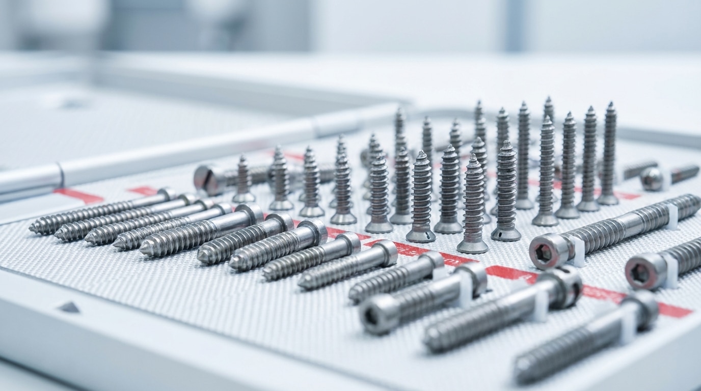
Before getting into specific types, it helps to understand the anatomy of an orthopedic screw. Every screw has the same basic components: a head (which interfaces with the plate or sits on the bone surface), a shaft (the smooth or threaded body), and a tip (self-tapping, self-drilling, or blunt/trocar). The thread is where the mechanical work happens — thread pitch (distance between threads), thread depth (height of the thread relative to the core diameter), and thread profile (buttress, V-type) determine how the screw grips bone.
Two measurements matter most: the outer diameter (the widest point of the thread) and the core diameter (the shaft without the thread). The ratio between these determines pullout strength and resistance to fatigue failure. A larger outer diameter relative to core diameter means deeper threads and more bone purchase. A larger core diameter means higher resistance to bending and shear forces on the screw itself.
The mechanical job of every orthopedic screw is the same: convert rotational torque into axial compression. When you drive a screw into bone, the threads grip the bone and the screw head presses the plate (or the near cortex) against the fracture, generating compression across the fracture surfaces. This is basic AO/ASIF principle — compression promotes primary bone healing.
Cortical Screws
Cortical screws are designed for purchase in the dense outer shell of bone — the cortex. They have a relatively small thread pitch (threads are closely spaced, typically 1.0-1.75mm apart depending on screw diameter) and shallow thread depth. This fine-pitch design maximizes the number of thread turns per unit length in dense cortical bone, which is where the holding power comes from.
Standard cortical screw diameters in orthopedic trauma include 2.0mm, 2.4mm, 2.7mm, 3.5mm, and 4.5mm. The 3.5mm cortical screw is the workhorse for upper extremity and small-bone fixation. The 4.5mm cortical screw is the standard for large-bone (femur, tibia) plating constructs.
Cortical screws are fully threaded — threads run the entire length of the shaft. When used in conventional (non-locking) plates, the screw head sits in the plate hole with a spherical undersurface that generates friction between the plate and bone. This friction is what holds the construct together. The screw compresses the plate against the bone surface, and the friction between plate and bone provides stability.
When Cortical Screws Are Used
- Plating of diaphyseal fractures in cortical bone (mid-shaft humerus, forearm, tibia)
- Lag screw technique through dense cortical bone (when the gliding hole is drilled to the outer diameter)
- Any application where the screw must purchase in thick, dense cortex
Cancellous Screws
Cancellous screws are engineered for the softer, trabecular bone found in metaphyseal and epiphyseal regions — the ends of long bones, vertebral bodies, and the pelvis. They have a wider thread pitch (threads are farther apart) and deeper threads compared to cortical screws. The wider pitch means fewer threads per unit length, but each thread captures more trabecular bone in its path. The deeper threads increase the surface area of bone-screw contact in a material that is inherently less dense than cortex.
Cancellous screws are commonly available in 4.0mm and 6.5mm diameters. The 6.5mm cancellous screw, with its large thread profile, is a standard choice for fixation in the proximal and distal femur, proximal tibia, and calcaneus.
Most cancellous screws are partially threaded — the threads occupy only a portion of the shaft (typically 16mm or 32mm of thread length on a 6.5mm screw). The unthreaded shaft acts as a gliding segment: the screw threads grip the far fragment, and as the screw head seats against the near fragment (or plate), it pulls the fragments together, generating interfragmentary compression. This makes the partially threaded cancellous screw a lag screw by design.
When Cancellous Screws Are Used
- Fixation of femoral neck fractures (6.5mm or 7.0mm cannulated cancellous screws)
- Fixation of tibial plateau fractures (subchondral support)
- Metaphyseal fixation in periarticular plating
- Syndesmotic fixation (3.5mm or 4.5mm tricortical screws, though technically cortical screws are also used here)
Locking Screws
Locking screws changed the mechanics of plate fixation fundamentally. Instead of relying on friction between the plate and bone (which requires the plate to be pressed tightly against the periosteum), a locking screw has a threaded head that engages threaded holes in the plate itself. The screw locks into the plate at a fixed angle, creating a construct where the plate and screws function as a single unit — an internal fixator.
This distinction matters clinically. In conventional plating, the construct fails when friction between the plate and bone is overcome — the plate slides on the bone, screws toggle in their holes, and fixation is lost. In locking plate constructs, each screw is a fixed-angle strut. The plate does not need to contact the bone surface at all to maintain stability. Loads are distributed across the entire construct rather than concentrated at individual screw-bone interfaces.
Locking screws are manufactured in the same diameter range as their conventional counterparts (2.4mm, 2.7mm, 3.5mm, 4.5mm, 5.0mm) and come in both cortical (fine pitch) and cancellous (wide pitch) thread profiles. The defining feature is always the threaded head — it may have a conical or cylindrical thread that matches the plate’s threaded hole.
When Locking Screws Matter Most
- Osteoporotic bone — the most important indication. In poor-quality bone, conventional screws lose purchase because the cortex cannot generate enough friction. Locking screws maintain their fixed-angle relationship with the plate regardless of bone density.
- Periarticular fractures — fractures near joints where the metaphyseal bone is thin and cancellous, and anatomic reduction is critical
- Bridge plating — spanning comminuted fractures where the plate must function as a load-bearing device across a zone of comminution without compressing the fracture
- Minimally invasive plate osteosynthesis (MIPO) — submuscular plate insertion where the plate cannot be compressed against the bone because access is limited
Cannulated Screws
Cannulated screws have a hollow core that allows placement over a guidewire. The surgeon places the guidewire first under fluoroscopic guidance, confirms position on imaging, and then drills and advances the screw over the wire. This technique provides two advantages: precise trajectory control (the wire acts as a rail) and the ability to confirm positioning before committing to the screw path.
Cannulated screws are available in nearly every diameter and can be either cortical or cancellous in thread design. The most common application is fixation of femoral neck fractures with three parallel 6.5mm or 7.0mm cannulated cancellous screws — a technique where guidewire positioning under fluoroscopy is critical to achieving proper spread pattern and avoiding joint penetration.
Other common cannulated screw applications include scaphoid fractures (headless cannulated screws), medial malleolus fixation, syndesmotic fixation, and small-fragment periarticular work where fluoroscopic-guided placement is preferred over direct visualization.
The tradeoff with cannulation is a smaller core diameter (the hollow center removes material), which slightly reduces the screw’s resistance to bending and fatigue failure compared to a solid screw of the same outer diameter. In practice, this is rarely clinically significant for standard applications.
Lag Screws and Interfragmentary Compression
A lag screw is not a specific screw type — it is a technique. Any screw can function as a lag screw if it generates compression across a fracture by gripping only the far fragment. The near fragment must be free to slide along the screw shaft as the head advances. There are two ways to achieve this:
- Partially threaded screws — the threads engage only the far fragment while the smooth shaft passes through the near fragment without gripping it. This is a lag screw by design.
- Lag technique with a fully threaded screw — the near cortex is over-drilled to the outer diameter of the screw threads (creating a gliding hole), so the threads only engage the far cortex. This is a lag screw by technique.
Lag screw fixation is an AO fundamental. It produces interfragmentary compression that converts a fracture into a mechanically stable unit, enabling primary bone healing without callus formation. Lag screws are used in isolation (simple fracture patterns) or through a plate (combined lag screw and neutralization plate construct).
The key surgical decision: the lag screw must be perpendicular to the fracture line for maximum compression, or perpendicular to the long axis of the bone for maximum resistance to shear forces. In oblique fractures, these are not the same angle, and the surgeon must decide which mechanical priority governs. For a comprehensive look at orthopedic implant types and applications, see our orthopedic surgical implants guide.
Specialty Screws: Headless, Herbert, and Suture Anchors
Headless Compression Screws
Headless compression screws (the Herbert screw is the original design) are fully buried beneath the articular surface, making them the standard for intra-articular fracture fixation where a protruding screw head would damage the opposing cartilage surface. The screw has differential thread pitch — the leading threads have a wider pitch than the trailing threads. As the screw advances, the leading threads pull the far fragment while the trailing threads push the near fragment, generating compression across the fracture without any screw head.
Primary applications include scaphoid fractures, osteochondral fractures, capitellum fractures, and talar body fractures — any intra-articular location where hardware prominence is unacceptable.
Suture Anchors
Suture anchors are technically screw-type devices designed to fix soft tissue (tendons, ligaments, labrum) to bone rather than bone to bone. They consist of a bone-engaging component (threaded screw, press-fit plug, or knotless design) loaded with high-strength suture or suture tape. Modern anchors are available in PEEK, biocomposite (resorbable), or titanium. They range from 1.3mm (for hand and wrist work) to 6.5mm (for shoulder and hip labral repair).
Plate Fundamentals
Orthopedic plates serve one or more of four mechanical functions, and understanding these functions determines plate selection:
- Compression — the plate generates active compression across a fracture (dynamic compression plate)
- Neutralization — the plate protects a lag screw construct from bending, rotational, and shear forces without generating compression itself
- Buttress — the plate prevents displacement of a fracture fragment under axial load (e.g., a tibial plateau buttress plate prevents the lateral plateau from collapsing under weight-bearing)
- Bridge — the plate spans a zone of comminution, maintaining length, alignment, and rotation while allowing indirect fracture healing through callus formation
The same physical plate can serve any of these functions depending on how the screws are placed and whether compression is applied. A 3.5mm LC-DCP used with a lag screw through the plate functions as a neutralization plate. The same plate applied with eccentric screw placement (using the spherical glide principle of the DCP hole) functions as a compression plate. Context determines function.
Dynamic Compression Plates (DCP and LC-DCP)
The Dynamic Compression Plate (DCP) is the original AO plate design for generating axial compression across a fracture using the geometry of the screw hole. The DCP hole has an inclined, ramped surface — when a screw is inserted eccentrically (offset toward the fracture), the spherical screw head slides down the ramp as it is tightened, translating the bone fragment horizontally toward the fracture. Placing eccentric screws on both sides of the fracture generates axial compression without a separate compression device.
The Limited Contact DCP (LC-DCP) refined this design by reducing the plate’s undersurface contact area with the periosteum. Scalloped undersurfaces preserve periosteal blood supply under the plate, reducing the biological cost of plate application. The LC-DCP also features a more symmetrical hole design (the combination hole or combi-hole) that allows the screw to be placed in compression mode, neutral mode, or at varying angles.
Standard DCP and LC-DCP plates are available in 2.7mm, 3.5mm (narrow and broad), and 4.5mm (narrow and broad) configurations, referring to the screw size they accept. Lengths range from 3-hole to 16-hole or longer, selected based on the fracture pattern and the required working length.
Locking Plates and Fixed-Angle Constructs
Locking plates accept locking screws that thread into the plate, creating a fixed-angle construct. Most modern locking plates are combination plates — each hole accepts either a conventional (non-locking) screw or a locking screw. This gives the surgeon the option to use conventional screws for compression (using the DCP glide principle) and locking screws for fixed-angle stability in the same construct.
Anatomically pre-contoured locking plates have become standard for periarticular fracture fixation. These plates are designed to match the surface anatomy of specific anatomic locations:
- Proximal humerus — locking plate contoured to the lateral proximal humerus with multiple fixed-angle locking screw options for the humeral head
- Distal radius — volar locking plate contoured to the volar surface of the distal radius with subchondral locking pegs or screws supporting the articular surface
- Proximal tibia — lateral and medial locking plates contoured to the tibial plateau with locking rafting screws for subchondral support
- Distal femur — lateral locking plate with multiple fixed-angle options for the condylar block
- Distal tibia (pilon) — medial, anterolateral, and posterolateral locking plates
- Calcaneus — lateral locking plate
These pre-contoured designs reduce intraoperative contouring time and improve the plate-bone fit, which is especially valuable in MIPO techniques where the plate is slid submuscularly and cannot be contoured under direct vision.
Reconstruction and Periarticular Plates
Reconstruction plates have notches cut between the screw holes that allow the plate to be bent and twisted in three dimensions. This malleability is essential for plating around complex anatomic surfaces — the pelvis (acetabular and pelvic ring fractures), the clavicle, the distal humerus, and other locations where a straight plate cannot conform to the bone surface.
The 3.5mm pelvic reconstruction plate is the standard for acetabular fracture fixation. It can be contoured along its length and across its width to follow the curved surfaces of the innominate bone, the quadrilateral surface, and the pelvic brim.
Periarticular-specific plates (not to be confused with anatomically pre-contoured locking plates) include designs for the olecranon, the distal humerus (parallel and perpendicular plating systems for bicolumnar fractures), the clavicle (superior and anterior contours), and the proximal femur (dynamic hip screw sideplate, blade plate).
Materials
Most orthopedic plates and screws are manufactured from 316L stainless steel or Ti-6Al-4V titanium alloy. Stainless steel is stiffer (higher modulus of elasticity), less expensive, and easier to contour intraoperatively. Titanium is more biocompatible, MRI-compatible (less artifact), and has a lower modulus closer to bone, which theoretically reduces stress shielding. Titanium is the standard for periarticular locking plates and any application where MRI surveillance will be needed postoperatively. Mixing titanium screws in stainless plates (or vice versa) causes galvanic corrosion and is never acceptable. For more details on how these devices are manufactured, see our guide on orthopedic implant manufacturing and quality testing.
Sourcing Orthopedic Screws and Plates
Orthopedic trauma is not elective. Fractures arrive in the emergency department on their own schedule, and the hardware to fix them needs to be available when the surgeon is ready to operate. This makes procurement speed and inventory depth the two most critical variables in choosing a hardware supplier.
A facility that routinely operates on distal femur fractures needs lateral distal femoral locking plates in multiple lengths, 5.0mm locking screws in every length from 30mm to 90mm, and conventional screws for the diaphyseal segment — all available in sterile trays the morning after the patient is admitted. A supplier that ships from a central warehouse with a 3-5 day lead time does not solve this problem.
SLR Medical Consulting supplies orthopedic screws, plates, and fixation hardware to surgical facilities nationwide from fully stocked warehouses with zero-lead-time processing. When the fracture shows up, the hardware shows up. Browse our orthopedic hardware catalog or place a surgical order to discuss your facility’s fixation needs.
Frequently Asked Questions About Orthopedic Screws and Plates
What is the difference between a cortical screw and a cancellous screw?
Cortical screws have fine-pitch, shallow threads designed for dense cortical bone. Cancellous screws have wide-pitch, deep threads designed for the softer trabecular bone found in metaphyseal and epiphyseal regions. Cancellous screws are often partially threaded to function as lag screws, while cortical screws are typically fully threaded. Using the wrong screw type in the wrong bone quality is one of the most common fixation errors — a cortical screw in soft cancellous bone will not achieve adequate purchase, and a cancellous screw in dense cortex will strip during insertion.
When should a locking plate be used instead of a conventional compression plate?
Locking plates are indicated when bone quality is poor (osteoporotic patients), when the fracture is periarticular (near a joint), when bridge plating across comminution is planned, or when MIPO technique is used and the plate cannot be compressed against the bone surface. In young patients with good bone quality and simple diaphyseal fracture patterns, conventional compression plating with standard cortical screws remains the gold standard. Many modern plates are combination designs that accept both conventional and locking screws, giving the surgeon flexibility to mix techniques within a single construct.
Can titanium and stainless steel implants be mixed in the same construct?
No. Mixing titanium and stainless steel in the same construct causes galvanic corrosion — an electrochemical reaction between dissimilar metals in a biological fluid environment. This accelerates corrosion at the metal interface, releases metallic ions into surrounding tissue, and can cause implant failure, tissue reaction, and pain. All screws in a plate must be the same material as the plate. This extends to washers, nuts, and any metallic component in contact with the construct.
What determines whether a plate functions as compression, neutralization, bridge, or buttress?
The function is determined by how the screws are placed and whether the fracture is compressed, not by the plate design itself. A DCP plate with eccentric screws generating axial compression is functioning as a compression plate. The same plate applied over a lag screw construct without active compression is a neutralization plate. The same plate spanning a comminuted zone without fracture reduction is a bridge plate. And if it is placed to resist axial collapse of a depressed fragment (e.g., lateral tibial plateau), it is a buttress plate. Understanding the mechanical function you need determines how you apply the hardware.
About SLR Medical Consulting: SLR Medical Consulting has been supplying surgical facilities nationwide for over a decade with orthopedic hardware, spine instrumentation, biologics, and sports medicine devices. Our zero-lead-time delivery model means your surgical schedule runs on your timeline, not your supply chain’s. Explore our hardware catalog or place a surgical order today.